|
|
|
Stem cell therapy for osteoarthritis
The following pictures are from the website of the National Institute of Arthritis and Musculoskeletal and Skin Diseases (NIAMS), part of the National Institutes of Health (NIH):
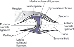
A Healthy Joint

In a healthy joint, the ends of bones are encased in smooth cartilage, which is protected by a joint capsule lined with a synovial membrane which in turn produces synovial fluid. The capsule and fluid protect the cartilage, the muscles, and the connective tissues.
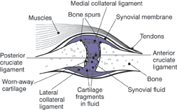
A Joint With Severe Osteoarthritis

With osteoarthritis, the cartilage becomes worn away. Spurs grow out from the edge of the bone, and synovial fluid increases. The joint therefore feels stiff and sore. From: NIAMS - National Institute of Arthritis and Musculoskeletal and Skin Diseases.
In the new field of "tissue engineering", stem cell therapy offers a novel form of treatment for patients suffering from osteoarthritis. Using the right type of stem cells, it is now possible to rebuild damaged cartilage.
Researchers at the Duke University Medical Center have "reprogrammed" adult stem cells by taking small deposits of fat from behind the kneecap and developing these fat cells into cartilage and bone cells which may then be grown into replacement tissue for people suffering with osteoarthritis. Cellular compatibility is guaranteed, and the possibility of immune rejection is eliminated since the stem cells are derived from a person's own tissue. The particular stem cells that are used in this procedure, namely, stromal cells derived from adipose tissue, are obtained using a minimally invasive approach. Scientists then treat the cells with a series of enzymes and, after centrifugal separation, the stromal cells are then further treated with a biochemical "cocktail" of steroids and growth factors which induces the cells to differentiate into multiple lineages. Adipose tissue has been shown to constitute a rich source of progenitor cells, and simply by controlling the biochemical environment in which the stem cells are grown, researchers have been able to grow different types of cells from these adult stem cells.
Laboratory stem cell studies are often conducted by "infusing" the stem cells into a three-dimensional matrix, which resembles most closely the internal "topography" of the human joint. Utilizing such a model, researchers at the University of Illinois at Chicago (UIC) have grown bone and cartilage from adult stem cells, in a study conducted in December of 2003, thereby successfully forming the ball structure of a joint that is found in the human jaw. The newly created joint, formed in the laboratory, exhibits the same characteristic shape and tissue composition of the human original after which it was patterned. The creation of this type of ball structure, known as an "articular condyle", marks an historic milestone in a process which could ultimately be used to regenerate knees, hips, and other joints that are typically damaged by osteoarthritis. Dr. Jeremy Mao, the director of the Tissue Engineering Laboratory at UIC, and an associate professor of bioengineering and orthodontics, has stated that, "This represents the first time a human-shaped articular condyle with both cartilage- and bone-like tissues was grown from a single population of adult stem cells. Our ultimate goal is to create a condyle that is biologically viable - a living tissue construct that integrates with existing bone and functions like the natural joint." Funded by multiple grants from the National Institutes of Health and the Whitaker Foundation, Dr. Mao used mesenchymal stem cells derived from the bone marrow of rats in the creation of this articular condyle. It has been shown that mesenchymal stem cells, which are present in a variety of types of adult tissue, are capable of differentiating into any kind of connective tissue, such as tendons, skeletal muscle, ligaments, cartilage, bone and teeth. With the addition of growth factors and other chemical substances, these stem cells may be "engineered" to produce whatever specific type of tissue is desired. Such custom-made tissues have been shown to replicate identically the characteristics of their naturally occurring counterparts, such as an extensive collage matrix with deposits of calcium salts, in the case of bone, and collagen with large amounts of proteoglycans, in the case of cartilage. While additional research is needed before tissue-engineered condyles are ready for the clinical treatment of patients suffering from osteoarthritis, rheumatoid arthritis, injuries or congenital anomalies, Dr. Mao nevertheless believes that, ultimately, the procedure could be adopted for total hip and knee replacements. "Our findings represent a proof-of-concept for further development of tissue-engineered condyles," Dr. Mao has said. (From the Journal of Dental Research). Adult bone marrow is a particularly rich source of stem cells in this new field of tissue engineering. As the inner, spongy mass that fills the core of long bones, bone marrow is easily accessible from such bones as the femur and tibia of the legs.
Similarly, in November of 2004, researchers at the Israel Institute of Technology, Technion, succeeded in building cartilage tissue in vivo, outside of the laboratory. Professor Joseph Mizrahi, chairman of Technion's Biomedical Engineering Department, together with Dr. Dror Seliktar invented a new type of bioreactor, which produces the mechanical stimulation that is necessary for building the tissue. As Dr. Mizrahi explains, "The basis is a polymer that serves as a scaffold within which we implant the cells. The polymer solidifies, thus enabling transfer of mechanical loads to the cells, which then begin to create protein. Eventually, this protein replaces the polymer scaffold which wears away." Dr. Seliktar adds that the "scaffold" is engineered to degrade during the dynamics of the protein production, since, "The scaffold becomes superfluous when the protein is created," he explains. The research was conducted in collaboration with Dr. Jennifer Elisseeff from The Johns Hopkins University in the Baltimore, MD, who succeeded in growing fetal stem cells in goat tissue in such a manner that bone cell development is simulated. "We want to direct the basic, primitive cell to develop into a cartilage cell or a bone cell," explains Prof. Mizrahi. "We are researching if existing environmental conditions cause the cell to become a cartilage or bone cell."
It is commonly understood that the naturally regenerative ability of cartilage tissue when damaged is extremely limited and virtually non-existent in adults. Several independent researchers have found that stem cells from muscle can also be coaxed to repair cartilage, however. As previously described, damage to articular cartilage (cartilage covering the ends of bones where they meet in a joint) is a frequent occurrence due to injury, illness and natural aging, and is a common cause of degenerative osteoarthritis. While conventional treatments are problematic, the new field of stem cell therapy has shown repeated success in the targeted repair of specific sites of damaged articular cartilage.
Numerous studies also indicate that muscle tissue constitutes a previously unrecognized source of stem cells that can be "reprogrammed" to develop into a variety of cells, including cartilage and bone cells. In a study published in the February 2006 issue of Arthritis and Rheumatism, researchers designed a study in which muscle-derived stem cells (MDSCs) were engineered with a therapeutic protein in order to repair articular cartilage defects in rats. The research was led by Johnny Huard, PhD, director of the Growth and Development Laboratory at Children's Hospital of Pittsburgh and an associate professor in the departments of Orthopaedic Surgery and Molecular Genetics and Biochemistry and Bioengineering at the University of Pittsburgh School of Medicine. The study involved the use of 12-week-old rats, demonstrating that skeletal muscle is a readily available and promising source of stem cells which can differentiate into cartilage. As Dr. Huard describes, "The MDSCs used here served as good carriers of a therapeutic gene and enabled the delivery of appropriate amounts of BMP-4 protein to the injury site." The findings show great promise in the repair of articular cartilage. (From, "Cartilage Repair Using Bone Morphogenetic Protein 4 and Muscle-Derived Stem Cells," Ryosuke Kuroda, Arvydas Usas, Seiji Kubo, Karin Corsi, Hairong Peng, Tim Rose, James Cummins, Freddie H. Fu, Johnny Huard, Arthritis and Rheumatism, February 2006; 54:2; pp. 433-442).
In an accompanying editorial in the same issue, Dr. Mary B. Goldring, of the New England Baptist Bone and Joint Institute and Harvard Medical School in Boston, Massachusetts, points out the significance of Dr. Huard's study as the first to investigate enriched stem cells from muscle in the treatment of damaged cartilage. Dr. Goldring also acknowledged that the study "provides proof-of-principle for performing MDSC implantation in cartilage of adult humans, since 12-week-old rats are considered to be young adults." Although other studies have suggested that the tissue lining the space between joints (the synovium) might also be a source of stem cells, obtaining these cells is more invasive than muscle biopsies. Dr. Goldring also notes that patients could potentially serve as their own donors, although she agrees that, "Further work is warranted to determine the chondroprogenitor potential of MDSCs in adult humans and their capacity to form cartilage in vivo." (From, "Are Bone Morphogenetic Proteins Effective Inducers of Cartilage Repair? Ex Vivo Transduction of Muscle-Derived Stem Cells," Mary B. Goldring, Arthritis and Rheumatism, February 2006; 54:2; pp. 387-389).
While stem cells derived from muscle represent some of the newer areas of research in cartilage repair, stem cells derived from fat, bone marrow and cord blood have long-established and well documented histories in the successful repair and regeneration of cartilage. The richest stem cell source of all, however, remains cord blood, which logically exhibits the greatest capacity for the repair and regeneration of any tissue.
With stem cell therapy, it is now possible for entire joints to be relined with new cartilage that has been bio-engineered to specific parameters. Sufferers of osteoarthritis may now regain their strength and mobility, free of pain, with newly developed methods of stem cell treatment.
According to the website of the National Institute of Arthritis and Musculoskeletal and Skin Diseases (NIAMS), a division of the National Institutes of Health (NIH),
"In 2004, NIAMS and other institutes and offices of the NIH began recruiting participants for the Osteoarthritis Initiative (OAI). The OAI is a collaboration that pools the funds and expertise of the NIH and industry to hasten the discovery of osteoarthritis biomarkers: physical signs or biological substances that indicate changes in bone or cartilage. Researchers are collecting images and specimens from approximately 5,000 people at high risk of having osteoarthritis as well as those at high risk of progression to severe osteoarthritis during the course of the study. Scientists are following participants for 5 years, collecting biological specimens (blood, urine, and DNA), images (X-rays and magnetic resonance imaging scans), and clinical data annually. For updates on this initiative, go to www.niams.nih.gov/ne/oi."
On August 1st of 2006, the NIH issued a press release informing the public that the first set of data from the OAI is now available. A description of the study may be viewed at the website of the Johns Hopkins Arthritis Center, and data on participants is accessible on the website of the University of California at San Francisco (UCSF).
Although The United Nations, the World Health Organization and 37 countries have proclaimed 2000 to 2010 as the "Bone and Joint Decade", individual countries have also proclaimed their own decades in which extra research would be dedicated to bone and joint health. In the United States, 2002 to 2011 was declared the "National Bone and Joint Decade." In proclaiming this Decade, President George W. Bush made the following declaration:
"Bones, joints and connective tissue are the structure upon which all other systems of the body depend. They give us strength, mobility, protection and stability. Our musculoskeletal structure is a complex system of tissue and bone that is regularly subjected to trauma, metabolic and genetic processes, and the gradual wear and tear of an active life. In the U.S., musculoskeletal disorders are a leading cause of physical disability. Such conditions also affect hundreds of millions of people around the world."
"The National Bone and Joint Decade, 2002-2011, envisions a series of international initiatives among physicians, health professionals, patients, and communities, working together to raise awareness about musculoskeletal disorders and promoting research and development into therapies, preventative measures, and cures for these disorders. Advances in the prevention, diagnosis, treatment, and research of musculoskeletal conditions will greatly enhance the quality of life of our aging population."
"The National Institutes of Health, the National Institute of Arthritis and Musculoskeletal and Skin Diseases, and other Federal agencies support many bone and joint studies. Industry and private professional and voluntary agencies support other initiatives. This work involves scientists examining the possible genetic causes of bone and joint diseases and studying how hormones, growth factors, and drugs regulate the skeleton. Other researchers are studying bone density, quality, and metabolism, and other ways to increase the longevity of joint replacements for those whose daily activities have become painful, difficult, or even impossible. These research efforts can help relieve pain and suffering and give countless children and adults the opportunity for a better life. Thanks to the hard work of these dedicated researchers, we have made great progress in understanding and treating musculoskeletal disorders. I commend their efforts and encourage them to pursue diligently further research that will help those suffering from these disorders. And I hope that all Americans will learn more about musculoskeletal problems, their long- and short-term effects, and the therapies and treatments available to help them."
From the website of the White House.
Musculoskeletal diseases such as osteoarthritis are recognized internationally not only as problems that impact individual lives, but also as problems that impact national economies. Such a global call for the effective treatment of these diseases represents an important political policy today, but it will acquire a tone of increasing urgency as the global population continues to age.
In the treatment of all forms of arthritis, especially osteoarthritis, stem cells offer a potentially useful therapy.
|
|
